


|
Yoshiko TAKAHASHI, Ph. D. 
 The
vertebrate body arises from a seemingly unremarkable clump of cells that
gradually organize into tissues displaying a wide variety of shapes and
functions. On closer observation, the individual cells that take part
in these morphogenetic processes also exhibit highly dynamic and varied
activity. The research in our lab concentrates on investigations into
the early stages of the body's development, when its structural plans
are formed and executed, as evidenced by the behavior of cells in our
primary model systems, the chicken and the mouse.
The
vertebrate body arises from a seemingly unremarkable clump of cells that
gradually organize into tissues displaying a wide variety of shapes and
functions. On closer observation, the individual cells that take part
in these morphogenetic processes also exhibit highly dynamic and varied
activity. The research in our lab concentrates on investigations into
the early stages of the body's development, when its structural plans
are formed and executed, as evidenced by the behavior of cells in our
primary model systems, the chicken and the mouse.
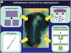
Visible patterns are defined by boundaries, but the means by which boundaries form in biological patterning processes is poorly understood. We study the formation of somites, embryonic regions that serve as precursors to much of the adult musculoskeletal system in vertebrates, seeking insights into the molecular underpinnings of boundary formation during that process. Previous work by our lab identified a boundary-inducing activity in inter-somite regions named the “segmenter”, which we have linked to Notch signaling and the activity of the Ephrin/Eph ligand-receptor complex.
Cells in the early embryo are mobile, migrating throughout the body as it takes shape, following sets of rules that direct their movements to specific destinations, allowing them to build functional complex structures, tissues and organs. We are studying how one such rule, which dictates that vascular development follows pathways laid down by neurons, operates in the establishment of a three-dimensional network formed by cells of the neural crest (precursors of the peripheral nervous system) and embryonic blood vessels through the study of cellular behaviors and the regulatory effects of inductive factors.
The physiological functions of many organs, such as lung and the lining of the gut, rely on the activity and organization of epithelial cells. In such organs, these cells form highly-ordered sheets, which in many cases roll up into tubular structures. Conversions of epithelial cells into mesenchymal cells, and vice-versa, collectively referred to epithelial-mesenchymal transitions (EMT), make the formation of even more complex structural arrangements possible. We are now investigating epithelial and tubular structures and the molecular mechanisms participating in EMT to clarify how these activities contribute to the building of the body.
