|
 |
Newt regeneration |
Regeneration of a newt's leg
(p-1.avi / 1008KB / 20秒) |
|
|
 |
 |
|
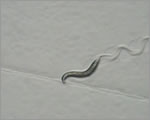
Planarian regeneration |
When a planarian is cut into pieces, each piece can regenerate into an individual anima
(p-2.avi / 484KB / 13秒) |
|
|
|
|
 |
 |
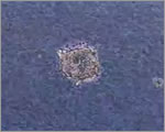
nou-darake gene |
When the function of the nou-darake gene is lost, brain cells grow throughout the planarian's body
(p-3.jpg / 152KB) |
|
|
 |
|
RNAi injection |
Injection of RNA molecules into the planarian body, used to "knock down" gene expression
(p-4.jpg / 164KB) |
|
|
|
|
 |
|
Planarian |
Wild type planarian
(p-5.jpg / 64KB) |
|
|
 |
|
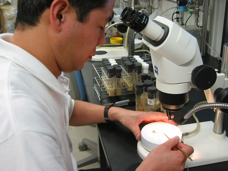
Nuclear transfer |
Nuclear transfer using a micromanipulator
(m-1.avi / 2.6MB / 41秒) |
|
|
|
|
 |
|
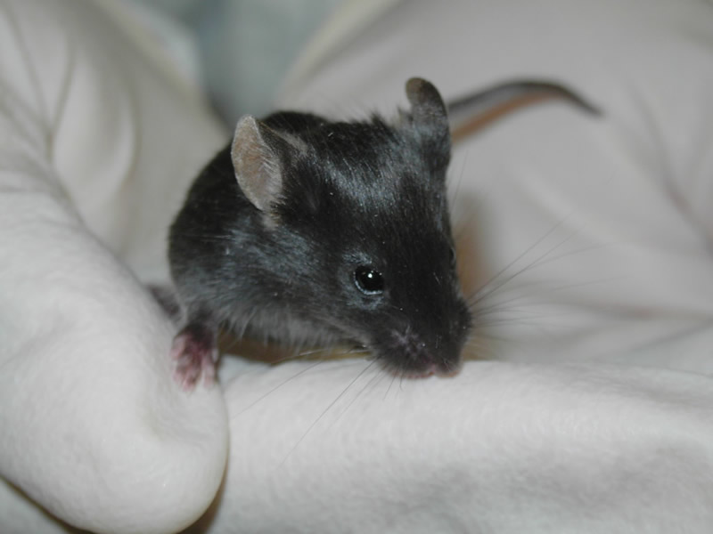
Cloned mice |
Mouse cloned using the nucleus from a tail-tip cell
(m-2.jpg / 164KB) |
|
|
 |
|
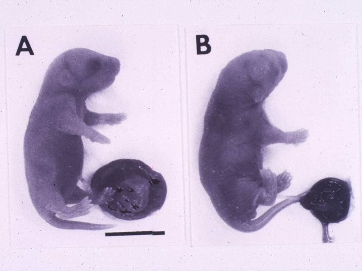
Defects of cloned mice 1 |
The cloned mouse embryo (A) has a larger placenta than normal (B)
(m-3.jpg / 208KB) |
|
|
|
|
 |
|
Defects of cloned mice 2 |
Cloned mice tend to obesity
(m-4.jpg / 212KB) |
|
|
 |
|
Knockout mice |
The deletion of a single gene results in a smaller phenotype(left) than wild type (right)
(m-5.jpg / 172KB) |
|
|
|
|
 |
|
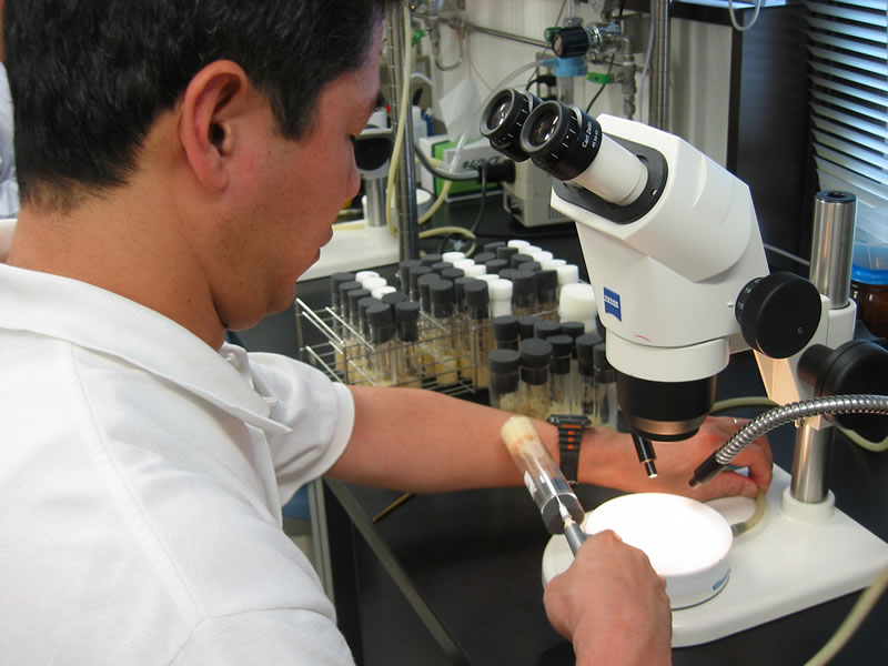
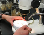
Micromanipulation |
The micropmanipulate makes it possible to work with cells and embryos under a microscope
(m-6.jpg / 196KB) |
|
|
 |
|
Drosophila experiment |
Anesthetizing fruit flies using carbon dioxide gas
(f-1.jpg / 196KB) |
|
|
|
|
 |
|
Drosophila experiment 2 |
Sorting male and female flies, and mutant phenotypes, under a microscope
(f-2.jpg / 196KB) |
|
|
 |
|
Drosophila mutant |
Mutation of a single gene (Antennapedia) causes legs to grow where antenna normally do
(f-3.jpg / 152KB) |
|
|
|
|
 |
|
Drosophila adult |
Normal (wild type) Drosophila adult
(f-4.jpg / 208KB) |
|
|
 |
|
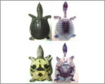
Turtle shell formation |
Bones corresponding to the ribs in other species form the shell in the turtle
(s-1.jpg / 200KB) |
|
|
|
|
 |
|
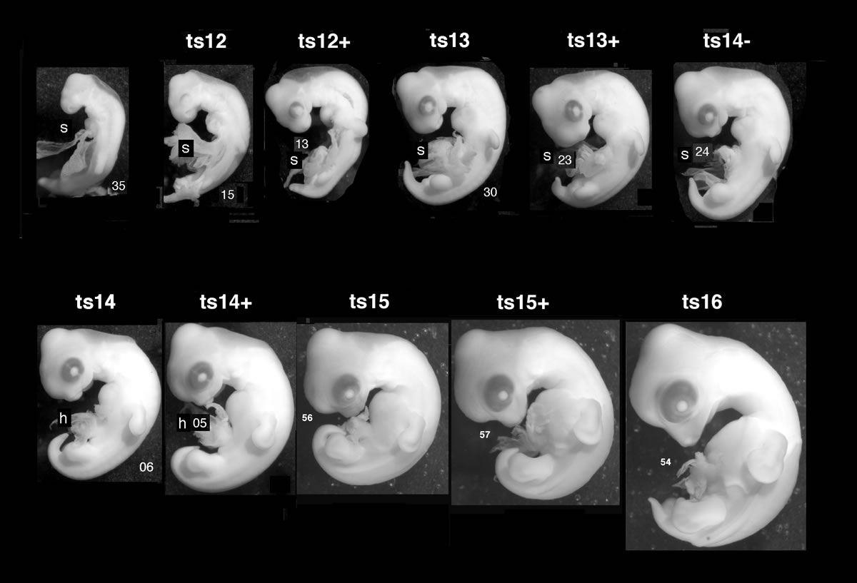
Turtle embryo |
Turtle embryonic development
(s-2.jpg / 164KB) |
|
|
 |
|
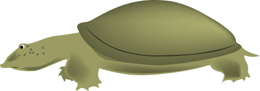
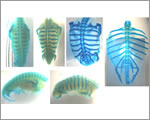
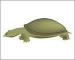
Chinese soft-shelled turtle |
Young Chinese soft-shelled turtle and its skeletal pattern
(s-4.jpg / 224KB) |
|
|
|
|
 |
|
Zebrafish skin pattern |
Skin pigment cells create the zebrafish's striped skin pattern
(z-1.jpg / 56KB) |
|
|
 |
|
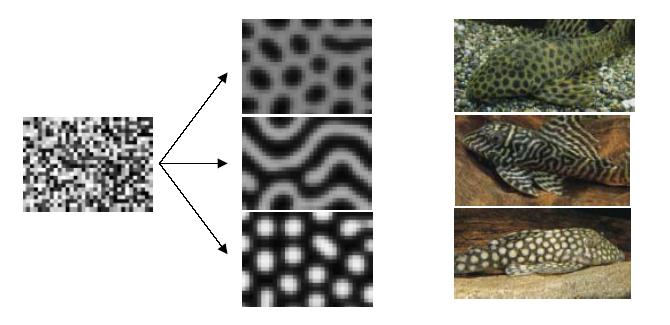
Three types of patterns seen on fish |
Simulation of generation of animal skin patterns (using the reaction-diffusion model)
(z-3.jpg / 92KB) |
|
|
|
|
 |
|
Angelfish |
Stripes on the skin of an angelfish
(z-4.jpg / 132KB) |
|
|
 |
|
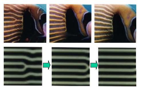
Changes in patterns |
Simulation of changes in skin pattern accompanying growth of the animal (reaction-diffusion model)
(z-5.jpg / 116KB) |
|
|
|
|
 |
|
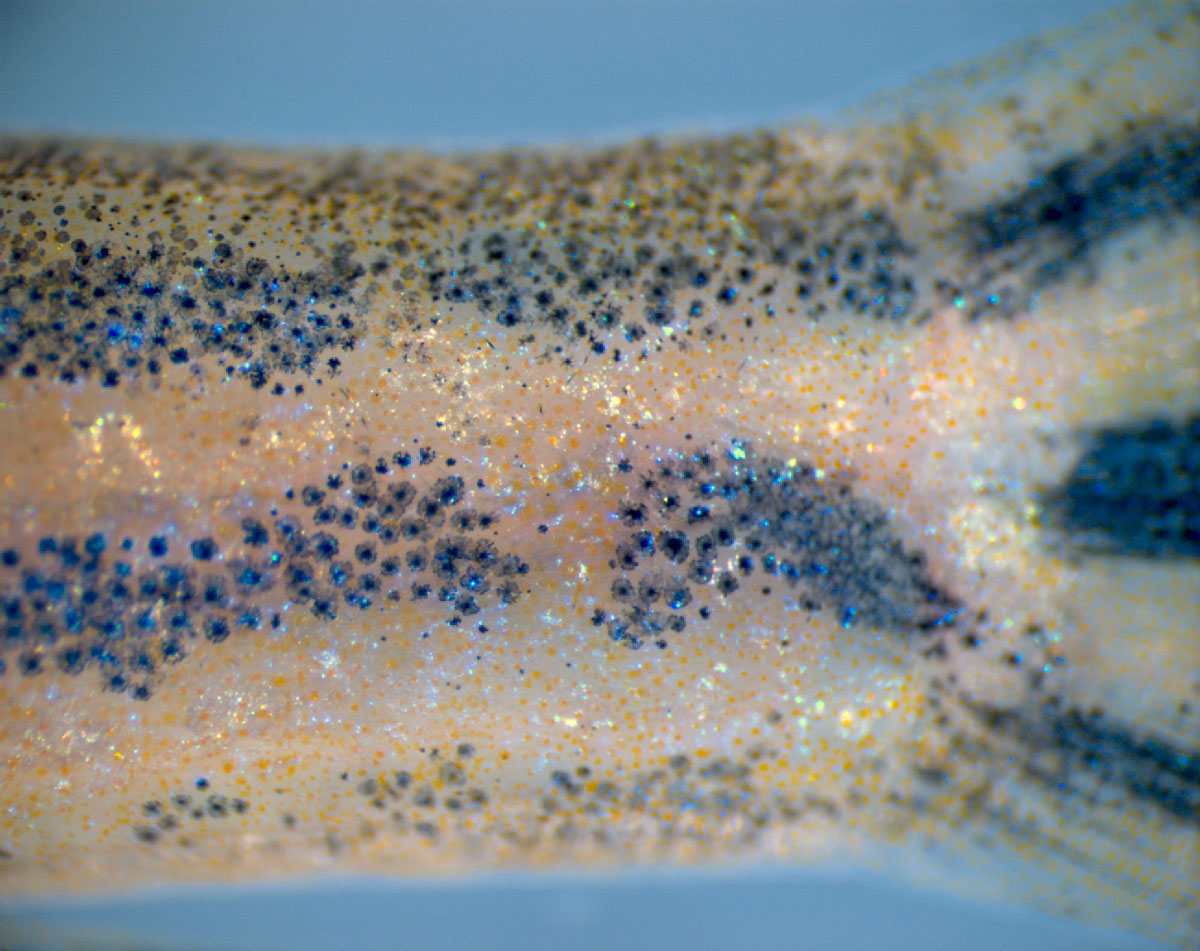
Reconstitution of zebrafish skin pattern (before) |
Pigment cells form the zebrafish skin pattern
(z-7.jpg / 420KB) |
|
|
 |
|
Reconstitution of zebrafish skin pattern (during) |
Destruction of pigment cells using a laser
(z-8.jpg / 364KB) |
|
|
|
|
 |
|
Reconstitution of zebrafish skin pattern (after) |
Reconstitution of patterns. Compare results of simulation and real skin pattern.
(z-9.jpg / 328KB) |
|
|
 |
|
Zebrafish |
Normal (wild type) zebrafish adult
(z-10.jpg / 88KB) |
|
|
|
|
 |
|
Sensory neurons |
Sensory neurons derived from primate ES cells
(a-1.jpg / 164KB) |
|
|
 |
|
Dopaminergic neurons |
Dopaminergic neurons derived from mouse ES cells
(a-2.jpg / 216KB) |
|
|
|
|
 |
|
Motor neurons |
Motor neurons derived from primate ES cells
(a-3.jpg / 132KB) |
|
|
 |
|
RNA injection 1 |
RNA injection into an early frog embryo under a microscope
(a-4.jpg / 200KB) |
|
|
|
|
 |
|
RNA injection 2 |
RNA injection into an early frog embryo under a microscope
(a-5.jpg / 180KB) |
|
|
 |
|
RNA injection 3 |
RNA injection into an early frog embryo under a microscope
(a-6.jpg / 136KB) |
|
|
|
|
 |
|
GFP tadpole |
GFP (fluorescent protein) used to identify the areas of expression of a target gene
(a-7jpg / 148KB) |
|
|
 |
|
GFP tadpole 2 |
GFP (fluorescent protein) used to identify the areas of expression of a target gene
(a-8.jpg / 132KB) |
|
|
|
|
 |
|
Tadpole (wild type) |
Normal (wild type) tadpole
(a-9.jpg / 100KB) |
|
|
 |
|
Tadpole (secondary embryo) |
Transfection of a certain gene causes development of a secondary embryo
(a-10.jpg / 108KB) |
|
|
|
|
 |
|
Animal cap |
The Xenopus animal cap is used to study induction factors
(a-11.jpg / 124KB) |
|
|
 |
|
Dorsal mesoderm |
Dorsal mesoderm induced by exogenous factors from the animal cap
(a-12.jpg / 124KB) |
|
|
|
|
 |
|
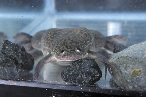
Xenopus laevis |
The African clawed frog (Xenopus laevis) is widely used in developmental biology research
(a-13.jpg / 200KB) |
|
|
 |
|
Roundworm embryogenesis |
Development of the roundworm, C. elegans.
(c-1.avi / 6.3MB / 66秒) |
|
|
|
|
 |
|
C. elegans (Wild type) |
Normal (wild type) roundworm adult
(c-3.avi / 5MB / 18秒) |
|
|
 |
|
C. elegans (Mutant1) |
Mutants for the rol-1 gene developmnt an abnormal cuticle (skin) and curl into coils
(c-4.avi / 3.6MB / 12秒) |
|
|
|
|
 |
|
C. elegans (Mutant2) |
Mutants for the gene unc-8 have abnormal nervous system function, and move in a amusing manner
(c-5.avi / 8.7MB / 32秒) |
|
|
 |
|
C. elegans (Mutant3) |
Mutants for the gene dpy-3 suffer abnormalities in cuticle (skin) development and are smaller than wild type worms
(c-6.avi / 4.9MB / 17秒) |
|
|
|
|
 |
|
C. elegans experiment |
Observation of C. elegans under a microscope
(c-7.jpg / 188KB) |
|
|
 |
|
C. elegans experiment 2 |
Selection of C. elegans under a microscope
(c-9.jpg / 200KB) |
|
|
|
|
 |
|
Chicken embryogenesis |
Chicken embryonic development
(ch-1.avi / 3.9MB / 29秒) |
|
|
 |
|
Chicken embryo experiment |
It is possible to observe and manipulate the chicken embryo through a hold made in the eggshell
(ch-2.jpg / 72KB) |
|
|
|
|
 |
|
Somitogenesis in chicken embryo |
Somites develop in pairs in a head-downwards direction
(ch-3.jpg / 80KB) |
|
|
 |
|
Gene transfection and embryo manipulation |
Somites expressing a target gene from one embryo are transplanted into a different embryo
(ch-4.jpg / 216KB) |
|
|
|
|
 |
|
Mouse ES cells |
Proliferating mouse ES cells
(e-1.avi / 3.1MB / 20秒) |
|
|
 |
|
Inducing differentiation of mouse ES cells |
Mouse ES cells that have been induced to differentiate into blood vessel cells
(e-2.avi / 5.5MB / 54秒) |
|
|
|
|
 |
|
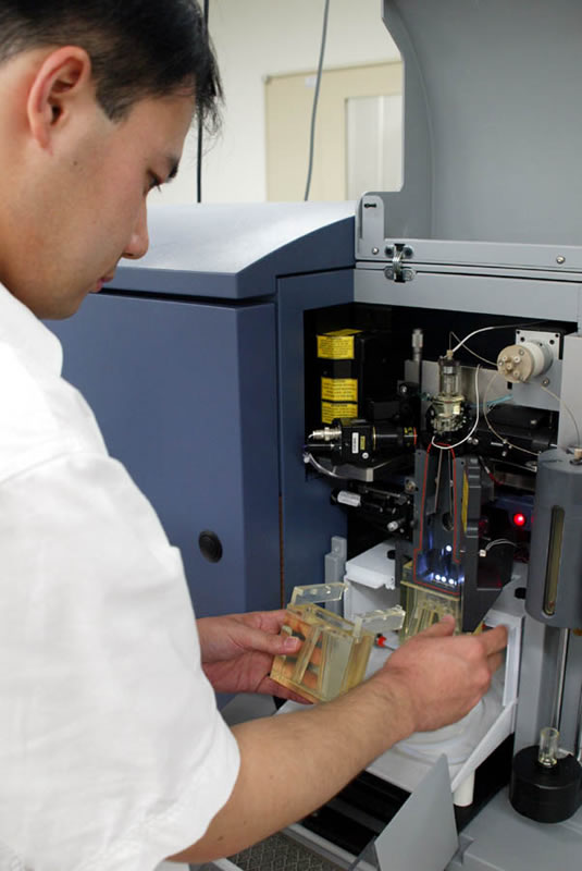
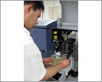
Cell sorter |
Cell sorters make it possible to isolate cells of a single type
(e-3.jpg / 160KB) |
|
|
 |
|
Cell experimentation lab |
Isolation of a single cell
(e-4.avi / 1.9MB / 10秒) |
|
|
|
|
 |
|
Fission yeast 1 |
Fission yeast is useful for the study of chromosome structure and the cell cycle
(y-1.jpg / 164KB) |
|
|
 |
|
Fission yeast 2 |
Fission yeast is useful for the study of chromosome structure and the cell cycle (the yeast shown here is fluorescence stained)
(y-2.jpg / 56KB) |
|
|
|
|
 |
 |
Newt 1 |
Newts are used to study regeneration
(i-1.jpg / 56KB) |
|
|
 |
|
Newt 2 |
Newts are used to study regeneration
(i-2.jpg / 164KB) |
|
|
|
|
 |
|
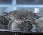
African clawed frog |
The African clawed frog has been used to study development for many years
(i-3.jpg / 84KB) |
|
|
 |
|
Chinese soft-shelled turtle |
The development of the turtle carapace is used as a model system at the CDB to study evolution
(i-4.jpg / 64KB) |
|
|
|
|
 |
|
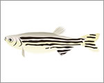
Zebrafish |
The zebrafish provides a good vertebrate model for use in mutant screens and analysis of knockdown phenotypes
(i-5.jpg / 68KB) |
|
|
 |
|
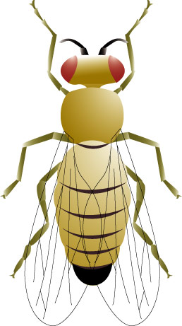
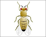
Drosophila |
The fruit fly Drosophila is widely used to study genetics
(i-6.jpg / 96KB) |
|
|
|
|
 |
|
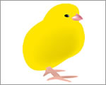
Chicken (chick) |
The chicken has a long history of use in the study of embryology
(i-7.jpg / 76KB) |
|
|
 |
|
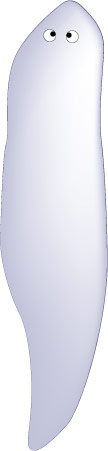
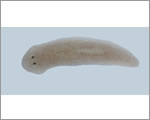
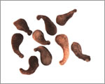
Planarian |
Planarians are used to study regeneration and stem cell biology
(i-8.jpg / 60KB) |
|
|
|
|
 |
|
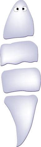
Planarian (section) |
When a planarian is cut into pieces, each piece can regenerate into a whole new animal
(i-9.jpg / 64KB) |
|
|
 |
|
Planarian (regeneration) |
When a planarian is cut into pieces, each piece can regenerate into a whole new animal
(i-10.jpg / 64KB) |
|
|
|
|
 |
|
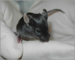
Mouse |
The mouse provides a mammalian model that is very similar in biological terms to the human and is used in many genetic studies
(i-11.jpg / 68KB) |
|
|
 |
|
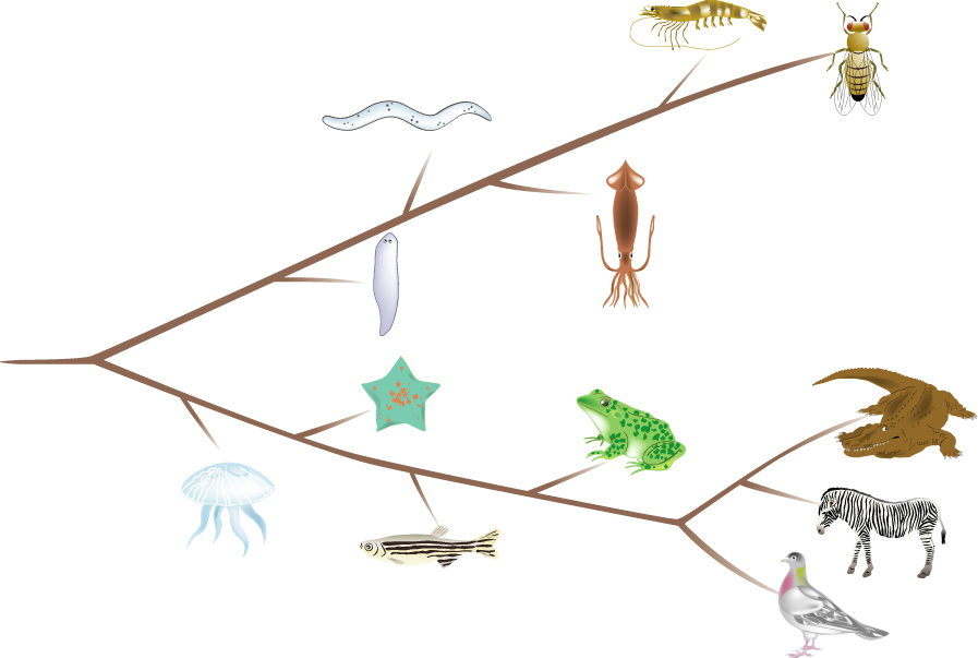
Phylogenetic tree |
Changes in developmentally important genes have led to the
(i-12.jpg / 184KB) |
|
|
|
|
 |
|
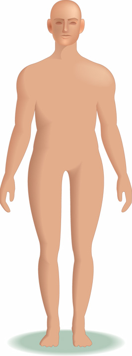
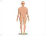
Human |
The 60 trillion cells that make up the human body share many features with those of other animals
(i-13.jpg / 80KB) |
|
|
 |
|
C. elegans |
Developmental processes can be observed directly in the transparent body of this 1mm long roundworm
(i-14.jpg / 48KB) |
|
|
|
|
 |
|
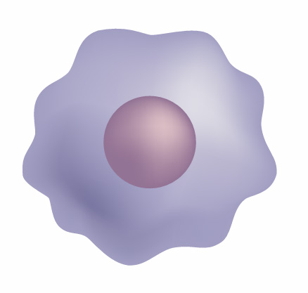
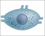
Cell
|
Structure of the animal cell
(i-15.jpg / 300KB) |
|
|
 |
|
DNA |
The DNA molecule carries genetic information encoded in sequences of nucleotides (A, G, T, C)
(i-16.jpg / 84KB) |
|
|
|
|
 |
|
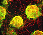
ES cells |
Embryonic stem (ES) cells can differentiate into any type of cell in the body
(i-17.jpg / 100KB) |
|
|
 |
|
Insulin-producing beta cells |
Insulin-producing beta cells have been derived from mouse ES cells; it is hoped that one day similar cells might be used to treat diabetes
(i-18.jpg / 84KB) |
|
|
|
|
 |
|
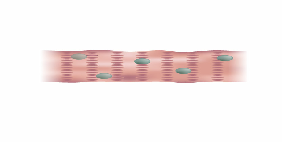
Muscle cells |
Heart muscles cells have also been derived from mouse ES cells
(i-19.jpg / 68KB) |
|
|
 |
|
Use of stem cells in medicine |
One day, embryonic and adult stem cells may be used to produce cells of specific types useful in treating human diseases
(i-20.jpg / 116KB) |
|
|
|
|
 |
|
Red blood cells |
Red blood cells carry oxygen to and waste products away from the body's other cells
(i-21.jpg / 72KB) |
|
|
 |
|
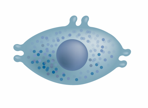
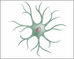
Neurons |
Different types of neurons can be generated from mouse ES cells
(i-22.jpg / 108KB) |
|
|
|
|
 |
|
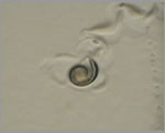
Fertilized egg |
Every human life began as a single fertlized egg
(i-23.jpg / 124KB) |
|
|
 |
|
Division of the egg (2-cell stage) |
The egg divides to produce more and more cells
(i-24.jpg / 128KB) |
|
|
|
|
 |
|
Division of the egg (4-cell stage) |
The egg divides to produce more and more cells
(i-25.jpg / 132KB) |
|
|
 |
|
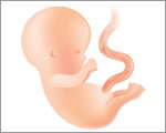
Embryo |
The embro develops through the processes of cell division and morphogenesis
(i-26.jpg / 112KB) |
|
|