News and Announcements from the CDB
The bending and stretching of sheets of epithelial cells into channels, tubes, and other topologies is a central process throughout much of development. In the pupa of Drosophila fruit flies, an interesting phenomenon occurs in which the cells of the male reproductive organ (terminalia) rotate consistently in the same direction with respect to the anterior-posterior axis, resulting in the wrapping of the spermiduct around the hindgut. In order for the terminalia to make its circuit, the surrounding epithelial cells must also move as a group in the same direction. But how they do this without a clear ‘conductor’ to coordinate their movements has been a mystery.
Now, a new report in Nature Communications from the Laboratory for Histogenetic Dynamics (Erina Kuranaga, Team Leader) working with the Laboratory for Physical Biology (led by Tatsuo Shibata) in the RIKEN Quantitative Biology Center (QBiC) shows how epithelial left-right asymmetry can give rise to such autonomous cellular movements.

Top: The pupal-stage terminalia makes a single clockwise rotation (movie).
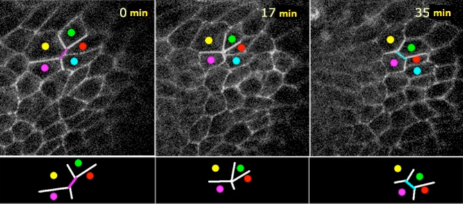
Bottom: Cells in adjacent epithelium show rightward-leaning positioning, as revealed by changes in the positions of cells marked by colored dots.
The pupal genitalia are located in the tail region, and their rotation is made possible by the coordinated movement of epithelial cells in abdominal segment eight (A8) that connect the terminalia with the abdomen. This A8 segment is divided into anterior (A8a) and posterior (A8p) components. Over 38 hours following the first 24 hours after puparium formation, first A8p makes a 180° turn, after which A8a completes the rotation by turning the remaining 180°. In this new study, the team began by isolating these tissues and observing them in culture to see whether they would retain their ability to rotate. They found that, even isolated from the rest of the abdomen, A8 epithelium still rotated autonomously.
They next sought to follow the changes in the positions of individual cells by labeling cell boundaries. While these cells maintained their cell-cell junctions, what the team found was that they constantly changed their borders and positions relative to each other throughout the rotation process. And intriguingly, the rearrangement of apical cell-cell boundaries showed a distinct rightward slant with respect to the A-P axis.
When the team examined the localization of the motor protein Myo-II, known to play a role in apical contraction, they found that while its distribution was not polarized asymmetrically within a cell prior to the start of rotation, its polarization showed a clear right-handed preference before and during the rotation of A8a. Higher concentrations of Myo-II were also associated with higher levels of left-right asymmetry, suggesting that the lopsided distribution of this factor might play a part in driving epithelium to move en masse.
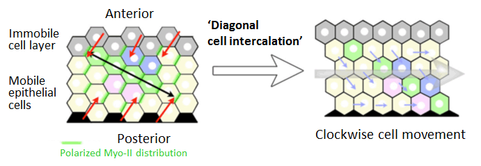
They looked at Myo-II in a fly mutant for Myo ID, in which the terminalia rotates opposite to the ordinary direction. In these cells, Myo-II tended to be more highly accumulated in left-leaning cell boundaries, again suggesting a role for this factor in coordinating epithelial movement at the group level. Finally, a mathematical simulation of the effects of left-right asymmetry gave further computational support to the notion that such rearrangements alone can give rise to autonomous movement by groups of cells, which can be made to be unidirectionally rightward when the cells are neighbored to the anterior by other immobile cells.

A model of unidirectional movement by left-right asymmetrical repositioning of cells. Accumulation of Myo-II at right-leaning cell boundaries results in
directional contraction and rearrangement of cell positions.
“These new results were made possible by combining advanced live imaging and quantitative modeling techniques,” says Kuranaga. “This finding of how left-right asymmetry can drive autonomous epithelial movements shows promise beyond the fruit fly, and may be of fundamental importance in a number of organogenetic processes in other species as well.”
| Link to article |
|---|